Links | About | Simon Moyes | Contact Us
- Surgeons site
- Ultrasound Scanner
- Treatment
- Historical Development
- Advantages and Contraindication
- Anatomy
- Instrumentation
- Theatre Layout
- Diagnostic Shoulder Arthroscopy
Quick Search
Latissimus dorsi transfer
Latissimus dorsi transfer has been proposed for the treatment of massive rotator cuff tear associated with severe functional impairment and chronic, disabling pain in young or active patients.
This technique substantially improves chronically painful, dysfunctional shoulders with irreparable rotator cuff tears, especially with intact subscapularis muscle. If subscapularis function is deficient it should be repaired if possible or if irrepairable other procedure as a reverse arthroplasty or arthrodesis are indicated.

The latissimus dorsi is a flat, broad muscle measuring about 20 by 40 cm. It extends from the posterior axilla to the midline of the back and inferiorly to the posterior portion of the iliac crest.
The latissimus functions mainly as an adductor and medial rotator of the arm. It also serves to pull the shoulder inferiorly and posteriorly.A candidate for a latissimus dorsi transfer could be a patient with:
- Preserved cartilage in the shoulder joint
- Massive rotator cuff tear
- Typical age 50-65 or active in work or sports activity
- Intact subscapularis tendon.
The operation is performed under general anaesthesia with an interscalene block for pain control. The patient is placed in the lateral decubitus position.
Before the tendon transfer an arthroscopy with intrarticular (sub-acromial decompression, A-C joint excision) or extrarticular procedures (Capsular release, bursectomy and axillary nerve isolation) is done on all patients in order to prepare the footprint for the tendon.
The posterior axillary fold is identified, and an incision is made just posterior to the lateral border of the latissimus dorsi, than skins flaps are elevated and the lateral and superior edges of the latissimus dorsi muscle are identified. The space between the latissimus dorsi and serratus anterior muscles is enlarged with blunt dissection. The neurovascular pedicle is preserved to its junction with the artery.
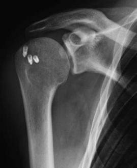
The posterior edge of the latissimus muscle is then freed and the tendon graft is harvested distally at his humeral insertion. The muscle is elevated and the flap after all the subcutaneous adhesion are freed can be rotated and transferred to the surgical great tuberosity. Once the flap is passed through the tunnel into the operative defect, it is insert and fixed with suture- anchor by arthroscopy without tension. The pedicle must be inspected to assure that no torsion or kinking of the vessels exists.
Because the transfer of the latissimus involves surgery around several important nerves and vessels, damage is possible.
Other complications include: bleeding, infection, continued shoulder pain, stiffness, seroma formation at the donor site lack of endurance of the arm in elevation and external rotation, failure to achieve successful result and need to redo the surgery, fracture, prolonged stiffness and or pain, implant failure, re-tear of the ligaments and complications relating to the anaesthesia.
