Links | About | Simon Moyes | Contact Us
- Surgeons site
- Ultrasound Scanner
- Treatment
- Historical Development
- Advantages and Contraindication
- Anatomy
- Instrumentation
- Theatre Layout
- Diagnostic Shoulder Arthroscopy
Quick Search
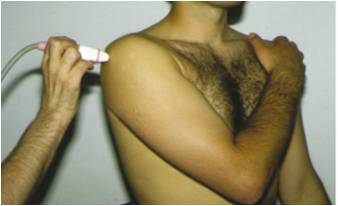
Ultrasound Scanner
Ultrasound (US) or sonography involves the sending of sound waves through the body. Those sound waves are reflected off the internal organs. The reflections are then interpreted by special instruments that subsequently create an image of anatomic parts. No ionizing radiation (x-ray) is involved in ultrasound imaging.
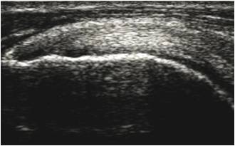
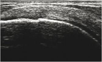
An ultrasound image is a useful way of examining the musculoskeletal system of the body to detect problems with muscles, tendons, joints and soft tissue. Ultrasound images are captured in real time, so they can often show movement, function, and anatomy, as well as enable radiologists to diagnose a variety of conditions and assess damage after an injury or illness.
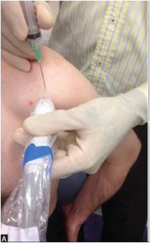
The Ultrasound Scans is a quick, safe and cheap procedure that allows the surgeons and the patients to have an immediate diagnosis and confirmation of many pathologies of the shoulder such as subacromial bursitis, calcifications, partial and complete rotator cuff tears and the arthritis of acromion-clavicular joint (ACJ).
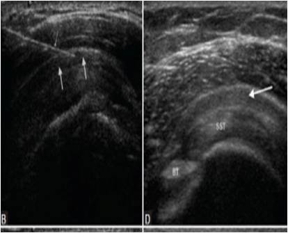
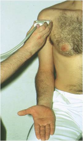
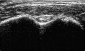
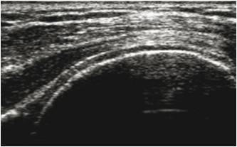
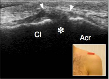
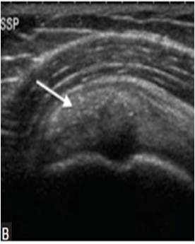
We use a standardized protocol to check each structures of the shoulder during US exam:
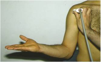
- Long Head Biceps TENDON: The patient sits with his arm in anatomical position, close to the body with the elbow 90° flexed and the dorsum of the hand resting on the patient’s upper leg.
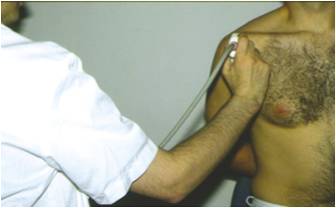
- Subscapularis Tendon: the patient has to exorotate his arm from the starting position outwards, while keeping his upper arm close to the trunk the elbow being still flexed at 90°
- Supraspinatus tendon: the patient put his hand on the back pocket
- Infraspinatus tendon: The arm is flexed and adducted with the hand resting on the contralateral shoulder.
- Acromion-clavicular joint (ACJ): in this case US is useful also to perform an inejction in this narrow joint.
- Calcifications
- Help for injections